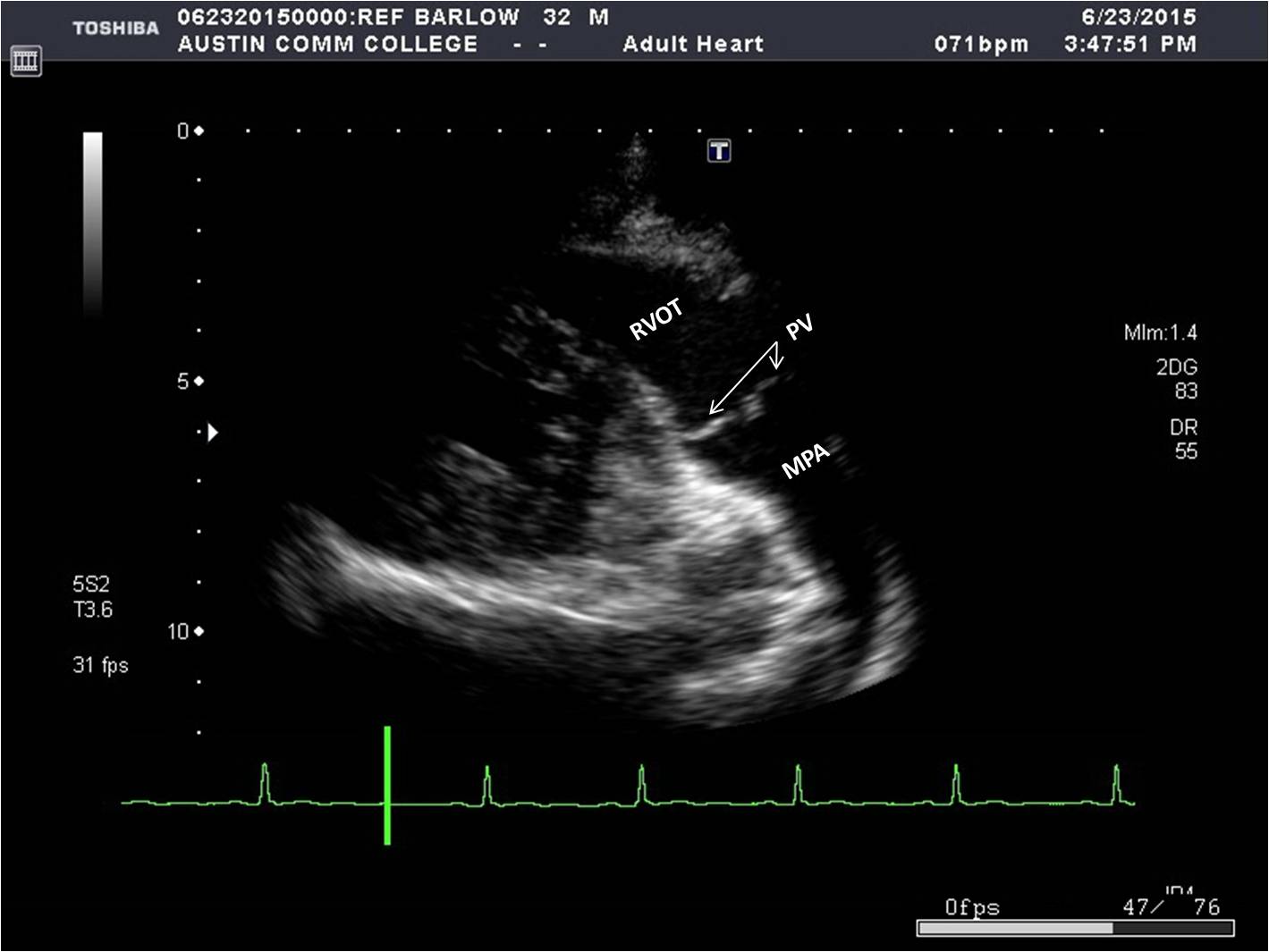
rvot on ultrasound


Parasternal Window | Sonography Resources

More pictures of rvot on ultrasound

Parasternal Window | Sonography Resources Parasternal Window | Sonography Resources A holistic sonographic view on congenital heart disease – How automatic  reconstruction using fetal intelligent navigation echocardiography (FINE)  eases the unveiling of abnormal cardiac anatomy part I: Right heart  anomalies - Weichert - Examples of ultrasound images of fetal heart in four-chamber (4CV),... |  Download Scientific Diagram Exploring the Pulmonary Vasculature with Korbin Haycock: RVOT Doppler.  FOAMed, FOAMcc, POCUS | thinking critical care 3.3.2 Right ventricular size | 123 Sonography PDF] Shape of the right ventricular outflow Doppler envelope and severity  of pulmonary hypertension. | Semantic Scholar Fetal Cardiac Screening Sonography – Radiology.World Guidelines for the Performance of the Sonographic Screening and  Echocardiography Examination of the Fetal Heart | Obgyn Key Fetal Echocardiography and the Routine Obstetric Sonogram Four-dimensional fetal echocardiography with spatiotemporal image  correlation (STIC): A systematic study of standard cardiac vie Figure 8 from CardioNet : Identification of fetal cardiac standard planes  from 2 D Ultrasound data | Semantic Scholar Fetal Echocardiography and the Routine Obstetric Sonogram Four-dimensional fetal echocardiography with spatiotemporal image  correlation (STIC): A systematic study of standard cardiac vie Diagnostics | Free Full-Text | Can Fetal Echocardiographic Measurements of  the Left Ventricular Outflow Tract Angle Detect Fetuses with Conotruncal  Cardiac Anomalies? Fetal Cardiac Screening Sonography – Radiology.World Basics of Fetal Echocardiography: Guidelines for Radiologists in India – JFR Parasternal Window | Sonography Resources Normal fetal cardiac anatomy. A, Sagittal reconstructed view of the... |  Download Scientific Diagram

4.1 299 votes
Article Rating